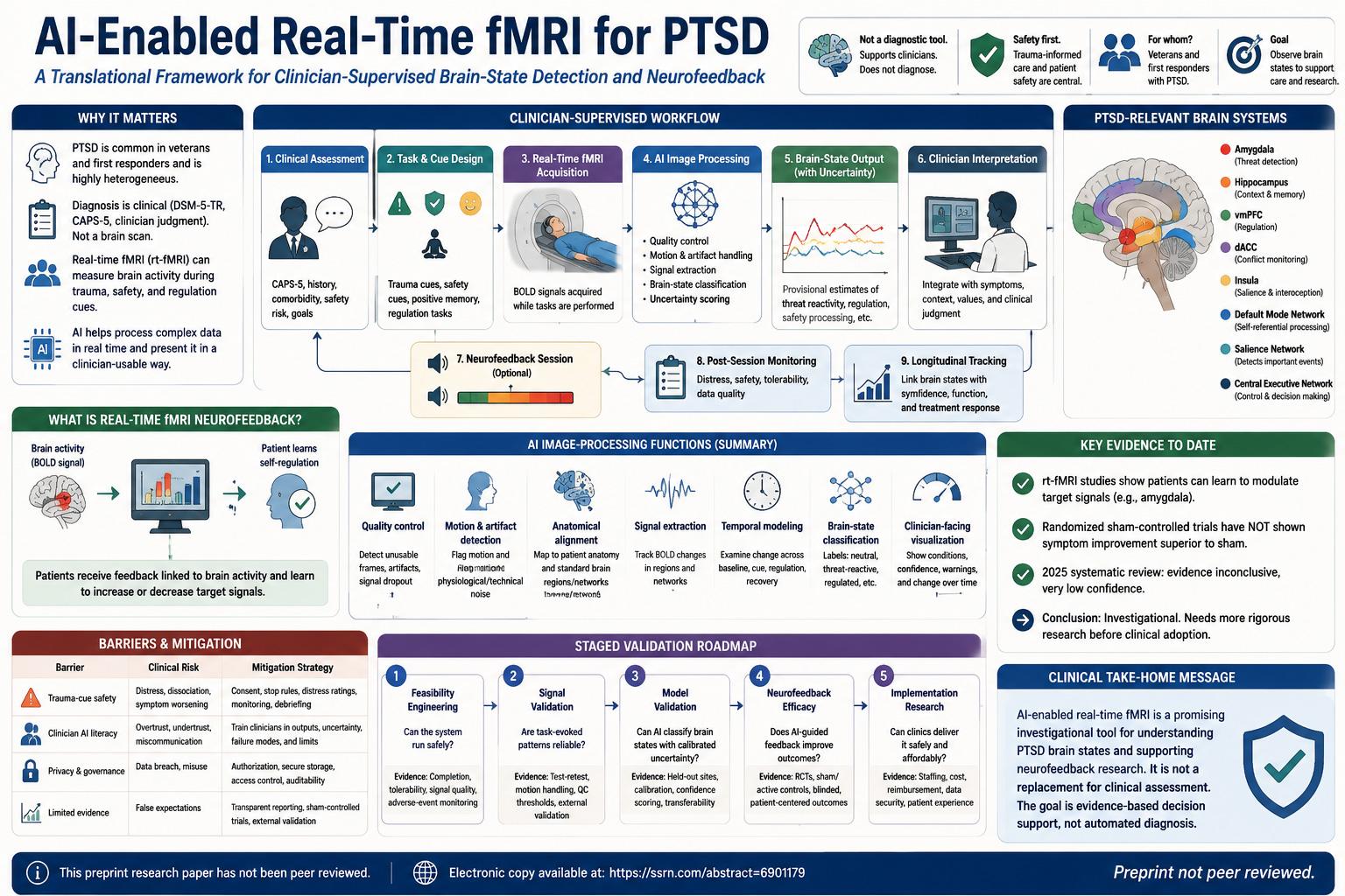
A non-peer-reviewed preprint article proposes a translational framework for using artificial intelligence with real-time functional MRI in posttraumatic stress disorder. The central idea is not that AI or fMRI should diagnose PTSD. Instead, the article argues that AI-enabled real-time fMRI may eventually support clinicians by detecting task-evoked brain-state patterns during trauma, safety, and regulation cues, helping select neurofeedback targets, accelerating interpretation, and integrating imaging outputs with structured clinical documentation.
The framework is deliberately cautious. PTSD diagnosis remains clinical and should continue to rely on DSM-5-TR criteria, structured assessments such as CAPS-5, clinician judgment, trauma history, symptoms, impairment, comorbidity, and safety evaluation. The proposed AI system would act as an image-processing and decision-support layer, not as an autonomous diagnostic authority. The paper emphasizes that real-time fMRI neurofeedback is still investigational for PTSD: existing trials show that some patients can learn to modulate target brain signals, but controlled evidence has not yet demonstrated symptom improvement superior to sham neurofeedback. The near-term goal is therefore evidence-based support for clinical understanding and research, not automated diagnosis or routine treatment replacement.
This paper addresses a difficult problem at the intersection of psychiatry, neuroimaging, artificial intelligence, and clinical translation. PTSD is diagnosed through clinical criteria and structured clinician assessment, not through a laboratory test or brain scan. That clinical foundation is essential because PTSD is not simply a single biological abnormality. It includes trauma exposure, intrusive memories, avoidance, negative changes in cognition and mood, altered arousal, functional impairment, comorbidities, and patient context. The author’s framework respects that reality rather than trying to replace it.
The paper’s starting point is PTSD heterogeneity. Two people can both meet diagnostic criteria for PTSD while having very different symptom patterns, cue responses, dissociation profiles, emotional regulation capacities, sleep problems, avoidance behavior, and treatment needs. The article notes that symptom-level criteria can produce more than 600,000 possible PTSD symptom combinations. This means a single diagnostic label can hide substantial biological and clinical variation. For veterans and first responders, the problem is even more complex because trauma may be repeated, occupationally embedded, and tied to duty, identity, and ongoing exposure risk.
The clinical question is therefore not whether fMRI can “see PTSD.” The article explicitly rejects that oversimplified idea. The more useful question is narrower: can real-time fMRI help clinicians observe brain-state changes during clinically meaningful tasks, such as trauma reminders, safety cues, positive memories, and regulation attempts? If it can, then imaging might eventually help answer questions that symptom interviews alone cannot fully resolve. For example: does a trauma cue trigger exaggerated threat reactivity? Does the patient recover toward baseline after the cue? Does a safety cue recruit regulatory networks? Can the patient learn to modulate a target signal during neurofeedback?
Real-time functional MRI, or rt-fMRI, is different from conventional post-session fMRI analysis. Traditional fMRI often analyzes data after the scan is complete. Real-time fMRI processes incoming blood oxygen level-dependent signals during scanning and can convert selected activity patterns into feedback. In neurofeedback, the patient receives a visual or auditory signal linked to a brain measure and tries to regulate it. In PTSD, prior studies have often focused on amygdala-related signals because the amygdala is involved in threat detection and emotional salience. However, the paper argues that PTSD-relevant brain states are unlikely to be reducible to one region.
This is scientifically important because fMRI does not read thoughts, memories, fear, guilt, trauma, or emotion directly. It measures BOLD signal changes, which are indirect, delayed vascular signals related to neural activity. A high or low signal in one region is meaningful only in the context of a task, timing, baseline, motion quality, and network interpretation. The paper repeatedly warns against treating a scan label as a diagnosis. The proposed system is best understood as a way to estimate task-linked patterns, not as a way to decode a person’s inner experience.
The author proposes a clinician-supervised workflow with four linked layers. First, the clinical-question layer: a psychiatrist or trauma clinician defines the reason for scanning. The question might involve cue reactivity, regulation capacity, safety-cue processing, neurofeedback target selection, or longitudinal monitoring. Second, the task-evoked imaging layer: the patient is exposed to neutral, trauma-related, safety, positive autobiographical, and regulation blocks during scanning. Third, the AI image-processing layer: AI performs real-time quality control, artifact handling, anatomical alignment, signal extraction, brain-state classification, uncertainty scoring, and visualization. Fourth, the clinical-interpretation layer: the clinician integrates the imaging output with symptoms, trauma history, distress, function, values, comorbidity, safety, and treatment response.
Figure 1 on page 4 provides the workflow visually. It moves from clinical assessment to task and cue design, real-time fMRI acquisition, AI image processing, brain-state estimate plus uncertainty, clinician interpretation, neurofeedback target selection, post-session monitoring, and longitudinal outcome tracking. The figure is simple, but it is central to the article’s argument because it shows the intended division of labor. The scanner captures data. AI processes the imaging stream. The clinician interprets the patient. That sequence is the ethical and clinical core of the framework.
The paper identifies PTSD-relevant systems including the amygdala, hippocampus, ventromedial prefrontal cortex, dorsal anterior cingulate cortex, insula, default mode network, salience network, and central executive network. These systems are linked to threat detection, contextual memory, self-referential processing, salience detection, regulation, and executive control. A clinically useful model, according to the paper, should therefore examine spatial patterns, temporal changes, and network relationships rather than a single “amygdala high” or “amygdala low” output.
That network-level view matters because PTSD is not just fear. Some patients may show strong threat reactivity. Others may show poor recovery after a cue ends. Others may show dissociation, altered self-referential processing, impaired safety learning, or difficulty recruiting prefrontal regulation. A single-region model risks flattening these differences. The proposed framework instead imagines labels such as neutral-like, threat-reactive, persistently dysregulated, successfully regulated, or low-confidence. These labels are not diagnoses. They are provisional task-defined brain-state estimates.
Table 1 on page 6 is one of the article’s most useful parts because it translates “AI” from a vague buzzword into concrete image-processing functions. The table lists image quality control, motion and artifact detection, anatomical alignment, region and network signal extraction, temporal feature modeling, brain-state classification, and clinician-facing visualization. Each function has a clinical or safety interpretation. For example, image quality control prevents poor data from becoming misleading clinical-looking output. Motion and artifact detection supports low-confidence outputs rather than forced classification. Brain-state classification produces provisional estimates for decision support, not diagnosis.
This table is important because real clinical harm can arise when AI output looks authoritative despite poor data quality. Trauma-related tasks may cause distress, movement, breathing changes, or physiological noise. Head motion can produce spurious connectivity patterns. Scanner artifacts and signal dropout can make a model appear to detect a brain state when it is really detecting unusable data. A responsible system must therefore be allowed to say “low confidence” or “uninterpretable,” and those warnings must be displayed as prominently as any classification label.
The article’s first underdeveloped function is accelerating and standardizing time-to-interpretation. In ordinary clinical practice, raw functional imaging data are not immediately useful to psychiatrists. The author argues that AI could reduce the time between scan acquisition and clinician-usable interpretation by automatically performing quality control, motion handling, alignment, feature extraction, and summary generation as the scan is being acquired. Instead of handing the clinician a stream of raw fMRI data, the system would present a curated, confidence-scored summary within or immediately after the session.
This speed is not valuable by itself. The author is careful to say that faster output can also spread error faster if the system is not validated. The value of speed depends on standardization. Functional neuroimaging can vary because of scanner differences, acquisition settings, preprocessing choices, motion thresholds, and operator decisions. Reproducible preprocessing pipelines and standardized data structures can reduce this variability. If a system labels a response as “threat-reactive,” the clinician needs to know that the same label means the same thing across sessions, sites, and patients as much as possible. Otherwise, longitudinal monitoring becomes unreliable.
The second underdeveloped function is integrating clinician documentation into a unified patient profile. This is one of the most clinically interesting parts of the paper. PTSD assessment is not only a set of imaging signals. It includes CAPS-5 severity, trauma narrative, avoidance patterns, dissociation, sleep, suicide risk, comorbidity, medication history, therapy history, and functional impairment. Much of that information lives in structured fields and free-text clinical notes. The paper suggests that AI could fuse text-derived clinical features with imaging-derived brain-state estimates to create a longitudinal patient profile.
This does not mean that AI should read notes and decide treatment alone. The proposed value is integration. A patient’s clinical record may show severe avoidance, trauma-cue distress, poor sleep, and dissociation. Their rt-fMRI session may show high salience-network reactivity, poor recovery after trauma cues, or weak recruitment of regulatory systems during safety cues. Integrated carefully, these two streams may help clinicians see convergences or discrepancies between subjective report, clinical observation, and task-evoked physiology. That could support more personalized neurofeedback target selection and research into treatment mechanisms.
The paper discusses natural language processing as one tool for extracting clinically meaningful constructs from psychiatric documentation. It also discusses multimodal fusion, which combines different types of data in a shared model. In the PTSD context, this could link structured assessment scores, clinician notes, task-evoked imaging patterns, and longitudinal outcomes. The promise is not a magical AI mind-reader. The promise is a better-organized, longitudinally updated clinical research profile that helps clinicians and investigators ask more precise questions.
Neurofeedback is the treatment-facing component of the framework. The patient receives feedback linked to a brain signal and attempts to regulate it. Candidate targets include amygdala reactivity during trauma recall, prefrontal regulation of threat response, posterior cingulate and default mode activity during self-referential processing, and broader salience or executive-control connectivity. The framework suggests that target choice should eventually be personalized rather than assigning every patient to the same region.
The article is notably cautious about the current evidence. Feasibility work in war veterans and combat veterans suggests that rt-fMRI neurofeedback can be tolerated and that some patients can learn to modulate target signals. However, controlled outcomes remain mixed. The paper highlights a randomized double-blind trial in which amygdala downregulation training improved control over amygdala activity but did not reduce PTSD symptoms more than sham. It also cites a 2025 systematic review judging controlled fMRI-neurofeedback evidence for PTSD inconclusive with very low confidence. This distinction is essential: learning to change a brain signal is not the same as achieving durable symptom improvement.
This is where many public discussions of neurotechnology become misleading. A brain-scan image, feedback graph, or AI confidence score can look persuasive. But the clinical question is not whether the patient can move a signal during a task. The clinical question is whether that learning improves distress, avoidance, sleep, functioning, quality of life, safety, and long-term recovery beyond placebo, sham, practice effects, nonspecific engagement, or standard care. The paper’s conservative stance protects readers from confusing mechanistic feasibility with proven treatment efficacy.
The workflow section describes how the process would work in practice. It begins with pre-scan assessment and safety screening. Patients should receive DSM-5-TR-informed evaluation with CAPS-5 or similar validated measures, trauma-history review, MRI safety screening, and risk assessment for suicide, dissociation, acute destabilization, intoxication, psychosis, severe claustrophobia, and unsafe implants. This is crucial because trauma cues during scanning can provoke distress or dissociation, and MRI itself has safety constraints.
Next comes task and stimulus design. The scan may include neutral stimuli, trauma-related cues or scripts, safety cues, positive autobiographical memory, and active regulation. AI may help organize or tag stimulus content, but clinicians must approve trauma-related materials. This is ethically important. Trauma cues are not generic images for many patients. They may be deeply personal, destabilizing, or clinically inappropriate depending on current risk and treatment phase.
During acquisition, the AI pipeline runs quality control, alignment, feature extraction, and classification. The system estimates whether the current pattern matches a task-defined target state. But the framework emphasizes that the system must be allowed to return “low confidence” or “uninterpretable.” This is a major safety principle. A forced label can be worse than no label if the underlying data are corrupted by motion, dropout, or poor task adherence.
After acquisition, the clinician reviews a concise summary: baseline response, peak trauma-cue response, recovery after the cue, response to safety cues, regulation success, image-quality concerns, and confidence level. That summary may guide the next neurofeedback run or inform treatment planning, but it does not override clinical assessment. Post-session monitoring then evaluates distress, tolerance, adverse effects, and whether the session produced usable data. Longitudinal tracking asks whether brain-state changes across sessions correspond to validated symptom and function measures.
Table 2 on pages 12 and 13 summarizes implementation barriers and mitigation strategies. The barriers include lack of a validated individual biomarker, BOLD delay and noise, limited neurofeedback evidence, model generalizability problems, privacy and governance challenges, trauma-cue safety, and clinician AI literacy. Each barrier is paired with a clinical risk and mitigation strategy. For example, the risk of overinterpreting scan output as proof of PTSD is mitigated by keeping diagnosis anchored in DSM-5-TR criteria and clinician judgment. The risk of distress or symptom worsening from trauma cues is mitigated by consent, stop rules, distress ratings, monitoring, and debriefing.
This table is valuable because it turns caution into operational design. It is not enough to say “AI should be used responsibly.” The framework specifies what responsibility means: uncertainty reporting, motion flags, external validation, privacy safeguards, access controls, clinician training, adverse-event monitoring, and clear limits on interpretation. In a PTSD context, these safeguards are not administrative extras. They are part of patient safety.
Privacy is especially important because this framework would combine trauma histories, clinical notes, psychiatric assessments, and neuroimaging outputs into a unified profile. That kind of dataset is highly sensitive. It may include protected health information, trauma narratives, suicide risk, occupational identity, imaging-derived markers, and longitudinal treatment responses. The paper therefore emphasizes authorization, secure storage, access control, auditability, de-identification where appropriate, and compliance with privacy and security rules. Data fusion increases clinical value but also magnifies privacy exposure.
Regulatory and reporting issues are also central. The article references AI medical imaging reporting frameworks such as CLAIM 2024, prediction model guidance such as TRIPOD+AI, clinical trial reporting extensions such as CONSORT-AI, and FDA clinical decision-support guidance. These standards matter because AI systems can fail in ways that are hidden unless data sources, preprocessing, model architecture, validation, calibration, uncertainty, and failure cases are transparently reported. In medical AI, a polished dashboard is not evidence. Transparent validation is evidence.
Table 3 on page 13 lays out a staged validation roadmap. The first stage is feasibility engineering: can the scanner, pipeline, dashboard, and feedback loop run safely during PTSD tasks? Required evidence includes completion, tolerability, workflow timing, signal quality, and adverse-event monitoring. The second stage is signal validation: are task-evoked patterns reproducible within and across patients? This requires test-retest reliability, motion handling, quality-control thresholds, and external validation. The third stage is model validation: can AI classify brain states with calibrated uncertainty? This requires held-out site testing, calibration, confidence scoring, and scanner transferability.
The fourth stage is neurofeedback efficacy: does AI-guided feedback improve regulation and outcomes? This requires randomized sham or active-control trials with blinded, patient-centered outcomes. The fifth stage is implementation research: can clinics deliver the workflow safely and affordably? This requires staffing, cost, reimbursement, data security, training, and patient-experience evaluation. This staged roadmap is one of the strongest parts of the paper because it prevents premature clinical adoption. It shows that a working prototype is only the beginning.
The paper’s limitations are partly the limitations of the field and partly the limitations of the article itself. The article is a conceptual translational review, not a systematic review, meta-analysis, device evaluation, or original patient-data study. It does not present a new rt-fMRI dataset, train a new AI model, validate a classifier, or test a clinical intervention. Its contribution is a framework: it organizes existing technologies and evidence into a clinician-supervised model. That makes the article useful as a roadmap, but not as proof that the proposed system works clinically.
Another limitation is that the clinical evidence for PTSD neurofeedback remains weak. The paper correctly acknowledges that controlled trials have not yet shown clear symptom benefit over sham. This means the framework’s clinical promise depends on future studies, not current routine readiness. A responsible article should not imply that veterans, first responders, or trauma survivors should currently receive AI-enabled rt-fMRI as standard PTSD care. The author avoids that overclaim.
There is also a deeper conceptual limitation. Brain-state patterns may not map neatly onto treatment selection. A patient’s scan response may vary depending on sleep, medication, dissociation, current stress, task design, scanner environment, trauma cue content, trust in the clinician, and even whether the patient feels safe inside the MRI scanner. A low response could mean low reactivity, dissociation, avoidance, task disengagement, medication effects, or poor signal quality. This is why clinician interpretation remains indispensable.
For veterans and first responders, the framework has special relevance but also special ethical demands. These populations may have repeated trauma exposure, moral injury, occupational identity issues, stigma concerns, and fears about job fitness or documentation. Any AI-generated brain-state profile must not become a simplistic label used to judge capability, responsibility, or credibility. The paper’s emphasis on decision support rather than automated diagnosis is therefore clinically and socially important.
The practical future of this framework would likely begin in academic medical centers, VA settings, DoD-linked research environments, or specialized trauma research programs. The infrastructure demands are high: MRI scanner time, real-time processing systems, trained imaging staff, trauma-informed clinicians, emergency protocols, secure data systems, and AI governance. This is not a low-cost near-term replacement for standard therapy. It is a research-grade adjunct that may eventually help personalize care if evidence develops.
The strongest idea in the paper is the division of labor between clinician and AI. AI is strongest at processing complex data streams quickly, flagging artifacts, standardizing pipelines, extracting patterns, and organizing longitudinal information. Clinicians are essential for understanding trauma context, diagnosis, risk, meaning, values, comorbidity, distress, and treatment planning. The framework is most credible when each side stays in its proper role.
The paper’s most important warning is that AI-enabled rt-fMRI should not be marketed or understood as a PTSD diagnostic machine. The article’s conclusions are clear: the component technologies make the workflow plausible today, but translation requires prospective, sham-controlled, externally validated studies with transparent reporting and patient-safety safeguards. The near-term goal is evidence-based decision support, not automated diagnosis.
For general readers, the meaning is this: brain imaging may eventually help clinicians observe how a patient’s brain responds to trauma cues, safety cues, and regulation efforts in real time. AI may help convert complex scanner data into a more usable clinical summary. But the science is not ready to replace clinical interviews, therapy, medication decisions, or human judgment. The value of this framework lies in careful integration, not technological overconfidence.
Overall, this preprint offers a thoughtful roadmap for a future research and clinical-support system. It respects the complexity of PTSD, acknowledges the limits of current neurofeedback evidence, and places AI in a supporting role. Its contribution is not a proven treatment or diagnostic tool; it is a disciplined proposal for how real-time fMRI, AI image processing, neurofeedback, and clinician documentation could be studied together without reducing trauma care to an algorithm.
Source and Method Note
Source title: AI-Enabled Real-Time fMRI for PTSD: A Translational Framework for Clinician-Supervised Brain-State Detection and Neurofeedback.
Author: Valeriana Colón, PhD.
Publication / preprint / report status: This is an SSRN-hosted preprint conceptual translational review / framework article. The PDF explicitly states that the manuscript has not been peer reviewed.
Peer-review status: Not peer reviewed. The findings and recommendations should be interpreted as non-peer-reviewed preprint evidence and conceptual analysis, not as validated clinical guidance.
Subject area: Posttraumatic stress disorder, real-time fMRI, AI-enabled medical imaging, neurofeedback, clinical decision support, veterans, first responders, trauma psychiatry, multimodal data fusion, and clinician-supervised brain-state interpretation.
Methods used: The article is a conceptual translational review. It presents no original patient data and is not a systematic review, meta-analysis, device evaluation, clinical trial, or stand-alone diagnostic AI proposal. Sources were identified through targeted searches of PubMed/MEDLINE, Google Scholar, federal guidance, and reporting standards related to PTSD, real-time fMRI, neurofeedback, amygdala, machine learning, multimodal fusion, natural language processing, clinical decision support, veterans, first responders, and AI medical imaging.
Framework structure: The proposed model includes four linked layers: a clinical-question layer, a task-evoked imaging layer, an AI image-processing layer, and a clinical-interpretation layer. The system is designed to support clinicians by processing real-time fMRI data, estimating task-defined brain states, displaying uncertainty, and integrating imaging output with clinical documentation. It does not diagnose PTSD.
Figures and tables: Figure 1 on page 4 shows the clinician-supervised AI-enabled rt-fMRI workflow, moving from clinical assessment through task design, real-time fMRI acquisition, AI image processing, uncertainty-scored brain-state estimation, clinician interpretation, neurofeedback target selection, post-session monitoring, and longitudinal outcome tracking. Table 1 on page 6 summarizes AI image-processing functions, including quality control, motion detection, anatomical alignment, signal extraction, temporal modeling, brain-state classification, and clinician-facing visualization. Table 2 on pages 12-13 lists implementation barriers, clinical risks, and mitigation strategies. Table 3 on page 13 provides a staged validation roadmap, from feasibility engineering to implementation research.
Formula and statistical explanation: The article does not introduce a new mathematical formula or original statistical model. It discusses BOLD signal interpretation, task-evoked brain-state classification, uncertainty scoring, multimodal data fusion, and validation standards. The proposed workflow depends on transparent preprocessing, quality control, calibration, external validation, and patient-centered clinical outcomes rather than a single diagnostic equation.
Important caution: This article is an explanatory interpretation of a non-peer-reviewed preprint. It is not medical advice, not a PTSD diagnosis, not a treatment recommendation, not a neurofeedback prescription, not a psychiatric device approval, not an MRI safety clearance, not an FDA approval, not a clinical protocol ready for routine care, not legal advice, not investment advice, not an engineering certification, not a religious ruling, and not an official policy order. PTSD assessment and treatment should remain under qualified clinical supervision using validated assessment tools, established therapies, patient safety procedures, and applicable medical and regulatory guidance.