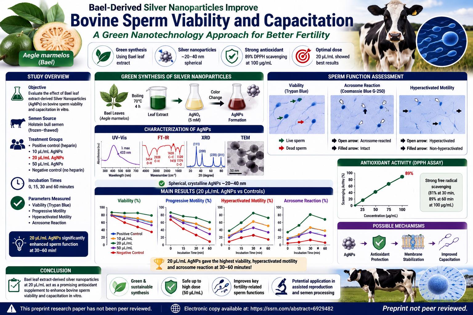
This study addresses a practical problem in veterinary reproductive biotechnology: how can bovine spermatozoa be supported during in vitro capacitation so that they remain viable, motile, and functionally ready for fertilization? In cattle breeding and assisted reproduction, sperm quality is not only about whether sperm cells are alive. They must also move effectively, undergo capacitation at the right time, develop hyperactivated motility, and complete the acrosome reaction in a coordinated way. If these steps happen poorly, too early, or too late, fertilization efficiency can decline.
Capacitation is a biological preparation process that spermatozoa must undergo before fertilizing an oocyte. During capacitation, the sperm plasma membrane changes: cholesterol is removed, membrane fluidity increases, calcium-related signaling changes, protein tyrosine phosphorylation occurs, and the sperm becomes capable of hyperactivated movement and acrosome reaction. Hyperactivated motility is a more vigorous, asymmetrical flagellar movement pattern that helps sperm penetrate the oocyte’s surrounding structures. The acrosome reaction is an exocytotic event in which the sperm releases enzymes from the acrosomal cap, helping it interact with and penetrate the zona pellucida.
The difficulty is that sperm capacitation is sensitive to oxidative balance. Moderate reactive oxygen species are not always harmful; small ROS levels participate in capacitation signaling. But excessive oxidative stress can damage sperm membranes, proteins, mitochondria, and DNA. Sperm cells are especially vulnerable because their membranes contain polyunsaturated fatty acids and because mature sperm have limited capacity for repair. This is why antioxidant strategies are frequently explored in semen preservation, sperm selection, and in vitro fertilization systems.
The authors propose Bael-derived silver nanoparticles as a possible antioxidant supplement for bovine sperm capacitation media. Aegle marmelos, or Bael, is a medicinal plant traditionally associated with antimicrobial, antidiabetic, anti-inflammatory, antioxidant, and other biological activities. The paper argues that Bael leaf extract contains phytochemicals such as phenolic compounds, flavonoids, alkaloids, tannins, sterols, and related metabolites that may help reduce silver ions into nanoparticles and stabilize the resulting nanomaterial. In this sense, the plant extract functions not merely as a raw material, but as a green synthesis agent.
The “green synthesis” concept is important. Conventional nanoparticle synthesis can involve harsh chemicals, expensive processes, and environmental concerns. Plant-mediated synthesis is attractive because extracts can act as reducing and capping agents under milder conditions. In the present study, Bael leaves were collected, washed, dried, ground, and extracted in water. Ten grams of leaf powder were mixed with 100 mL of double-distilled water and heated to 70°C for four hours. The filtered extract was then added to a 5 mM silver nitrate solution. The solution’s color change indicated reduction of silver ions into silver nanoparticles.
The study’s first experimental layer was nanoparticle characterization. This step matters because biological effects cannot be interpreted properly unless the material is reasonably described. Silver nanoparticles of different size, shape, surface chemistry, aggregation state, and crystallinity can behave differently in biological systems. A nanoparticle preparation that improves sperm parameters at one size range might be ineffective or toxic if particle size, coating, or concentration changes.
Figure 1 on page 12 shows the UV–Vis absorption spectrum of the Bael-derived silver nanoparticles. The maximum absorption peak was reported at 433 nm. This peak is interpreted as evidence of silver nanoparticle formation because silver nanoparticles commonly show surface plasmon resonance in this visible-light region. The exact peak position can shift depending on particle size, shape, aggregation, and local chemical environment. In simple terms, the UV–Vis result supports that silver ions were reduced into nanoscale silver structures rather than remaining only as dissolved ions.
Figure 2 on page 14 shows the FT-IR spectrum. The authors report a broad peak at 3454 cm-1, attributed to O–H stretching vibrations from phenolic compounds; a sharp peak at 1628 cm-1, attributed to C=C stretching; a peak at 2928 cm-1, attributed to aldehydic C–H stretching; and additional peaks around 1413 cm-1, 1120 cm-1, and 1079 cm-1, linked to C–H and C–O groups from phenolic acids, polysaccharides, carboxylic acids, esters, ethers, proteins, or metabolites. The key interpretation is that plant-derived biomolecules likely participated in both reduction and stabilization of the nanoparticles.
Figure 3 on page 15 shows the X-ray diffraction pattern. The diffraction peaks at approximately 38.1°, 44.26°, 64.44°, and 77.36° correspond to the (111), (200), (220), and (311) planes of a face-centered cubic silver lattice. The stronger intensity of the (111) plane suggests preferential crystallographic orientation. Using the Debye–Scherrer formula, the authors calculated an average crystallite size of about 20 nm. This supports the conclusion that the synthesized particles were crystalline silver nanoparticles.
Figure 4 on page 17 presents TEM and SAED evidence. The TEM images show mostly spherical nanoparticles, described as monodisperse and non-agglomerated, with an average crystallite diameter of 20–25 nm. The SAED pattern shows bright circular rings, consistent with crystallinity and matching the face-centered cubic structure indicated by XRD. These images are useful because they visually support the size and morphology claims. However, the paper also states in the abstract and highlights that the particles were mainly 30–40 nm. This creates a reporting inconsistency: the abstract suggests 30–40 nm, while the XRD and TEM sections report approximately 20 nm or 20–25 nm. A careful reader should not ignore this difference. It does not invalidate the study, but it means nanoparticle size reporting should be clarified before reproduction or application.
The antioxidant activity was evaluated using the DPPH assay. DPPH is a stable free radical that changes color when reduced by an antioxidant. The study calculated scavenging activity using the formula: DPPH scavenging activity (%) = [(Abs0 − Abs1) / Abs0] × 100, where Abs0 is the absorbance of the DPPH control and Abs1 is the absorbance in the presence of nanoparticles. Figure 8 on page 23 shows the antioxidant assay spectra. The reported antioxidant activity was 81.31% after 30 minutes and 89% after one hour at 100 µg/mL, suggesting strong free-radical scavenging capacity under the assay conditions.
This antioxidant result is relevant because oxidative stress is a major constraint in sperm handling. If the Bael-derived nanoparticles reduce excess ROS without suppressing physiological ROS signaling entirely, they could help preserve membrane integrity and functional capacitation. However, DPPH is a chemical antioxidant assay, not a direct measurement of ROS inside sperm cells. It shows radical-scavenging potential in a simplified assay system. It does not prove the exact intracellular mechanism by which sperm viability or capacitation changed.
The second experimental layer tested bovine spermatozoa. The study used frozen semen straws from Holstein bulls, thawed at 37°C for 30 seconds. Motile sperm were selected using a swim-up technique, a common method that allows more motile spermatozoa to move into the upper medium fraction. Sperm were incubated in SP-TALP capacitation medium and then evaluated for viability, progressive motility, hyperactivated motility, and acrosome reaction. The study compared a positive control containing sperm cells, heparin, and no nanoparticles; nanoparticle treatments; and a negative control described as sperm cells and nanoparticles only without heparin.
The paper’s methods section contains a notable inconsistency in treatment dose reporting. It describes five treatment groups: positive control, 10 µL nanoparticles, 20 µL nanoparticles, 100 µL nanoparticles, and negative control. However, the abstract, highlights, result discussion, and Table 1 report a high-dose treatment of 50 µL/mL rather than 100 µL. The table columns list Positive Control, 10, 20, 50, and Negative Control. Because the numerical results are presented for 50 µL/mL, this article interprets the reported data using 50 µL/mL while noting that the methods section should be corrected or clarified.
There is also a timing inconsistency. The methods discuss evaluation at 30, 60, and 90 minutes, while Table 1 reports values at 0, 15, 30, and 60 minutes. The narrative states that the 20 µL/mL treatment performed best at 30–60 minutes. This inconsistency matters because capacitation is time-dependent. A 15-minute, 30-minute, 60-minute, and 90-minute profile would lead to different biological interpretations. The study’s main pattern remains visible in the reported table, but future peer review should require clearer timing documentation.
Table 1 on pages 21–22 is the central data table for sperm outcomes. At 0 minutes, all groups had the same baseline values: viability 75.63 ± 0.44%, progressive motility 60.64 ± 0.48%, hyperactivated motility 0.00 ± 0.00%, and acrosome reaction 0.00 ± 0.00%. This baseline equality is important because it suggests the groups started from the same measured condition before incubation effects became visible.
At 15 minutes, the 20 µL/mL treatment showed the highest viability among the listed groups at 70.63 ± 0.44%, compared with 65.63 ± 0.44% in the positive control, 66.63 ± 0.44% at 10 µL/mL, 64.63 ± 0.44% at 50 µL/mL, and 50.63 ± 0.44% in the negative control. Hyperactivated motility was also highest in the 20 µL/mL group at 25.75 ± 0.25%, compared with 14.50 ± 0.26% in the positive control and 15.50 ± 0.24% in the negative control. Acrosome reaction followed a similar pattern: 20.75 ± 0.25% at 20 µL/mL versus 10.50 ± 0.26% in the positive control and 10.50 ± 0.24% in the negative control.
At 30 minutes, the 20 µL/mL group again showed the strongest functional profile. Viability was 65.63 ± 0.44%, higher than the positive control at 50.63 ± 0.44%, 10 µL/mL at 55.63 ± 0.44%, 50 µL/mL at 60.63 ± 0.44%, and the negative control at 30.63 ± 0.44%. Hyperactivated motility reached 34.68 ± 0.28% in the 20 µL/mL group, compared with 20.31 ± 0.25% in the positive control, 30.38 ± 0.35% at 10 µL/mL, 26.00 ± 0.27% at 50 µL/mL, and 10.50 ± 0.24% in the negative control. Acrosome reaction was also highest at 20 µL/mL, reaching 29.81 ± 0.23%.
At 60 minutes, sperm parameters declined in all groups, which is expected because sperm viability and motility often decrease with incubation time. However, the 20 µL/mL treatment still retained the best or near-best profile. Viability was 50.63 ± 0.44%, compared with 30.63 ± 0.44% in the positive control and 20.63 ± 0.44% in the negative control. Hyperactivated motility was 29.81 ± 0.22%, higher than the positive control at 15.31 ± 0.25% and the negative control at 8.50 ± 0.24%. Acrosome reaction was 20.81 ± 0.23%, again higher than the positive control and negative control.
The strongest practical interpretation of Table 1 is that 20 µL/mL was the most favorable concentration in this dataset. It preserved viability better over time and promoted functional capacitation indicators. The 10 µL/mL dose showed some improvement but was generally weaker. The 50 µL/mL dose did not provide additional benefit and sometimes performed below 20 µL/mL. This suggests a dose-response pattern with an optimal middle dose rather than a simple “more nanoparticles is better” relationship.
Progressive motility requires more careful interpretation. At 15 minutes, the positive control had the highest progressive motility value at 50.69 ± 0.72%, while the 20 µL/mL group had 40.81 ± 0.37%. At 30 minutes, the positive control still had 35.25 ± 0.73%, while 20 µL/mL had 30.00 ± 0.49%. At 60 minutes, the positive control had 27.06 ± 0.85%, while the 20 µL/mL treatment had 25.69 ± 0.64%. Therefore, the claim that 20 µL/mL consistently improved progressive motility over all controls is not fully supported by the table if progressive motility is considered alone. The stronger evidence for 20 µL/mL lies in viability, hyperactivated motility, and acrosome reaction, while progressive motility appears more complex.
This distinction is biologically meaningful. During capacitation, sperm motility changes from progressive forward movement toward hyperactivated movement. Hyperactivated sperm may not always look more progressively motile because their flagellar beat becomes asymmetrical and vigorous. Therefore, a decline in progressive motility does not automatically mean functional failure if hyperactivated motility and acrosome reaction increase appropriately. The key is whether the sperm are transitioning into a fertilization-competent state rather than simply moving forward in a straight line.
Figure 5 on page 18 shows Coomassie Blue G-250 staining used to distinguish acrosome-intact from acrosome-reacted spermatozoa. Acrosome-intact sperm display a dark blue apical ridge along the anterior head, while acrosome-reacted sperm lack this dark apical ridge and show lighter staining in the acrosomal region. This figure is important because it shows how the acrosome reaction was visually classified. It is not a decorative microscope image; it explains the biological endpoint behind the AR percentages in Table 1.
Figure 6 on page 20 shows hyperactivated motile bovine spermatozoa under the microscope. Hyperactivated sperm are identified by asymmetrical flagellar bends and large bent tails, while non-hyperactivated sperm have straighter tails. This visual distinction matters because hyperactivation is not merely “fast movement.” It is a characteristic movement pattern associated with capacitation and fertilization readiness. The figure helps readers understand what the HAM percentage represents.
Figure 7 on page 21 shows bovine spermatozoa stained with Trypan Blue after capacitation. Live sperm lack dark head staining and appear more lightly stained, while dead sperm show prominent dark blue head staining. This provides the basis for viability assessment. Trypan Blue is a membrane-integrity-based viability stain: cells with compromised membranes take up the dye, while viable cells exclude it. This is useful but limited; it measures membrane integrity, not DNA quality, mitochondrial function, or actual fertilizing ability.
The study’s conclusion states that bovine spermatozoa incubated with 20 µL/mL Bael nanoparticles for one hour showed significantly enhanced viability, progressive motility, hyperactivated motility, and acrosome reaction at 30–60 minutes compared with other treatments and controls. The table strongly supports improved viability, HAM, and AR at 20 µL/mL, but progressive motility should be interpreted more cautiously because the positive control had higher PM values at several time points. This is a good example of why detailed table reading is essential: the overall biological story can be promising while one parameter does not follow the broad claim perfectly.
The possible mechanism is antioxidant protection combined with improved capacitation environment. Bael-derived phytochemicals on or around the nanoparticles may scavenge free radicals, reduce lipid peroxidation, protect the sperm plasma membrane, and preserve viability. At the same time, the nanoparticles may influence sperm membrane dynamics or signaling in ways that support hyperactivation and acrosome reaction. However, the study did not directly measure ROS levels in the sperm medium, lipid peroxidation markers, mitochondrial membrane potential, ATP, calcium influx, protein tyrosine phosphorylation, DNA fragmentation, or fertilization rate in a controlled IVF endpoint. Therefore, the mechanism remains inferred rather than proven.
Safety is another key issue. The abstract states that AgNPs did not appear harmful or toxic to bovine spermatozoa up to 50 µL/mL under the in vitro conditions used. The table supports that 50 µL/mL was not catastrophic: viability at 60 minutes was 47.63 ± 0.44%, much higher than the negative control at 20.63 ± 0.44% and higher than the positive control at 30.63 ± 0.44%. However, “not toxic in this in vitro assay” is not the same as “safe in vivo.” Nanoparticles can behave differently in reproductive tracts, embryos, tissues, immune systems, and environmental settings. Silver nanoparticles in particular require careful toxicological evaluation because silver can be biologically active and dose-dependent.
The study’s strengths include its combination of green nanoparticle synthesis, multiple characterization techniques, antioxidant testing, and sperm functional assays. The use of UV–Vis, FT-IR, XRD, TEM, and SAED creates a reasonably broad material characterization profile. The sperm assays cover several functionally relevant endpoints rather than only viability. The dose comparison also helps identify an apparent optimum rather than assuming any nanoparticle exposure is beneficial.
The limitations are equally important. First, the study is a preprint and has not been peer reviewed. Second, the reporting contains inconsistencies in nanoparticle size, treatment dose, and incubation timing. Third, the experimental sample structure is not fully described in enough detail for strong reproducibility. The table states data are mean ± SE with n = 20, but the biological source of replicates, number of bulls, number of straws, and independence of observations should be clearer. Fourth, the study uses frozen-thawed Holstein bull semen in vitro, so results may not generalize to fresh semen, different breeds, different bulls, or in vivo reproductive conditions.
Fifth, the study does not report fertilization outcomes in a fully detailed way despite suggesting improved fertilization potential. It measures capacitation-related endpoints, but it does not provide a complete IVF embryo development dataset such as fertilization rate, cleavage rate, blastocyst rate, embryo quality, or pregnancy outcomes. Sixth, nanoparticle uptake, localization, dissolution, ion release, and interaction with sperm membranes were not measured. Seventh, long-term effects on embryos, oocytes, offspring, reproductive tract tissues, and environmental safety were not assessed.
Another limitation concerns terminology. The paper uses AgNPs, BNPs, and Bael nanoparticles in ways that can be confusing. The nanoparticles appear to be silver nanoparticles synthesized using Bael leaf extract, not purely plant-polysaccharide nanoparticles. The term BNPs may therefore be interpreted as Bael-derived silver nanoparticles. Clear terminology matters because a reader may otherwise think the particles are plant nanoparticles rather than silver nanomaterials capped or reduced by plant extract.
For assisted reproductive technologies, the study’s practical implication is promising but early. If replicated, a 20 µL/mL Bael-derived AgNP supplement might help maintain sperm viability and support capacitation in defined media. This could be relevant to bovine IVF, semen handling, sperm selection, or capacitation optimization. But before any practical use, the system would need stronger validation, including nanoparticle batch consistency, exact dose standardization, toxicity testing, ROS and lipid peroxidation measurements, IVF outcomes, embryo development, and regulatory evaluation.
For nanomedicine and reproductive biology, the broader lesson is that nanoparticles can have a narrow beneficial window. At low or moderate concentrations, antioxidant nanomaterials may support sperm function. At higher concentrations, they may offer no added benefit and may eventually become harmful depending on material and dose. The present study’s 20 µL/mL result fits this general principle: the best biological response came from a middle dose, not the highest dose.
For animal breeding and veterinary practice, the study should not be interpreted as a ready-to-use fertility treatment. It is not a semen extender protocol for farms, not an artificial insemination recommendation, and not proof that Bael-derived silver nanoparticles improve pregnancy rates. It is an in vitro preprint study showing that selected sperm function markers improved under specific laboratory conditions. Its value is scientific: it identifies a candidate additive and a plausible dose range that deserve more rigorous testing.
The most convincing result is the repeated advantage of 20 µL/mL for viability, hyperactivated motility, and acrosome reaction in Table 1. The most mechanistically interesting result is the strong DPPH antioxidant activity. The weakest points are the reporting inconsistencies and lack of direct mechanistic sperm oxidative-stress measurements. The most responsible conclusion is that Bael-derived silver nanoparticles may be useful as antioxidant-assisted capacitation media additives, but their reproductive safety and efficacy remain unproven beyond the reported in vitro setting.
Overall, this preprint contributes to the growing field of reproductive nanobiotechnology by linking plant-mediated nanoparticle synthesis with bovine sperm capacitation. It suggests that green-synthesized silver nanoparticles can influence sperm functional physiology in a concentration-dependent way. The study should not be overstated, but it offers a clear research direction: define the material precisely, clarify the dosing, test the mechanism directly, and evaluate whether improved capacitation markers translate into better fertilization and embryo outcomes.
Source and Method Note
Source title: Impact of Aegle marmelos Derived Silver Nanoparticles on Viability and Capacitation of Bovine Spermatozoa.
Authors: Iftkhar Ahmad, Asma Fatima, Md Aamir Aftab, Saber Abd-Allah, and Saiqa Ikram.
Publication / preprint / report status: This is an SSRN-hosted preprint experimental research manuscript in veterinary reproductive biotechnology and nanomaterials. The PDF explicitly states that the manuscript has not been peer reviewed.
Peer-review status: Not peer reviewed. The findings should be interpreted as non-peer-reviewed preprint evidence and require cautious reading, independent replication, and peer-review evaluation.
Subject area: Bovine sperm capacitation, veterinary assisted reproduction, green synthesis of silver nanoparticles, Aegle marmelos leaf extract, antioxidant nanomaterials, sperm viability, hyperactivated motility, acrosome reaction, and reproductive nanobiotechnology.
Methods used: The study prepared aqueous Bael leaf extract, synthesized silver nanoparticles by reducing silver nitrate with the plant extract, and characterized the nanoparticles using UV–Vis spectroscopy, FT-IR spectroscopy, X-ray diffraction, transmission electron microscopy, and selected-area electron diffraction. Antioxidant activity was assessed using the DPPH radical scavenging assay. Frozen-thawed Holstein bull spermatozoa were processed with a swim-up method and incubated in capacitation-supporting media with control conditions and nanoparticle treatments. Sperm viability, progressive motility, hyperactivated motility, and acrosome reaction were evaluated across incubation times.
Dataset and experimental structure: The sperm experiment used frozen Holstein bull semen straws, swim-up selection, SP-TALP capacitation medium, and treatment groups including a positive control, 10 µL/mL nanoparticles, 20 µL/mL nanoparticles, a higher nanoparticle dose reported in the table as 50 µL/mL, and a negative control. Table data are reported as mean ± SE with n = 20. The manuscript contains internal inconsistencies: the methods mention 100 µL treatment and 30, 60, and 90 minute evaluation, while the main result table reports 50 µL/mL and time points of 0, 15, 30, and 60 minutes. These details should be clarified before practical use or replication.
Figures, tables, and page numbers: Figure 1 on page 12 shows the UV–Vis absorption spectrum with a maximum around 433 nm, supporting AgNP formation. Figure 2 on page 14 shows FT-IR peaks indicating functional groups from Bael extract involved in reduction and stabilization. Figure 3 on page 15 shows the XRD pattern with silver FCC peaks and an estimated crystallite size of about 20 nm. Figure 4 on page 17 shows TEM and SAED analysis, indicating mostly spherical, crystalline, monodisperse nanoparticles with reported 20–25 nm diameter. Figure 5 on page 18 shows Coomassie Blue G-250 staining distinguishing acrosome-intact and acrosome-reacted spermatozoa. Figure 6 on page 20 shows hyperactivated and non-hyperactivated bovine sperm motility patterns. Figure 7 on page 21 shows Trypan Blue staining for live and dead spermatozoa. Table 1 on pages 21–22 reports sperm viability, progressive motility, hyperactivated motility, and acrosome reaction across treatments and incubation times. Figure 8 on page 23 shows DPPH antioxidant assay spectra and supports the reported antioxidant activity.
Formula and statistical explanation: The DPPH scavenging activity formula is: DPPH scavenging activity (%) = [(Abs0 − Abs1) / Abs0] × 100. The Debye–Scherrer equation was used to estimate nanoparticle crystallite size from XRD peak broadening: d = 0.89λ / βcosθ. Sperm outcome data were analyzed using one-way ANOVA with Duncan’s test, and hyperactivation motility and acrosome reaction were analyzed using Kruskal–Wallis one-way analysis of variance. Statistical significance was set at P < 0.05.
Important caution: This article is an explanatory interpretation of a non-peer-reviewed preprint. It is not veterinary medical advice, not a fertility-treatment recommendation, not a semen-processing protocol ready for field use, not an IVF clinical instruction, not a livestock breeding directive, not a nanoparticle safety certification, not regulatory approval, not legal advice, not investment advice, not an engineering certification, not a religious ruling, and not an official policy order. Any use of nanoparticles in reproductive media should require qualified veterinary, reproductive, toxicological, and regulatory review, especially because in vitro sperm findings do not automatically establish in vivo reproductive safety or fertility benefit.