Deborah DeSilets’ paper asks a deceptively simple question: what happens when biological systems that evolved to process information through slow, resistant, chemical, and spatially constrained media are placed inside digital environments that operate almost without friction? To answer this, the manuscript builds a bridge between developmental biology, neuroscience, and digital design theory. Its main argument is not that embryos and brains are the same system, but that both depend on a shared physical logic: information becomes meaningful only when it travels through a medium that shapes, slows, attenuates, and structures it.
The paper’s first major problem is biological spatial intelligence. In developmental biology, the classic question is how an embryo knows where to form different body parts. Every cell in an organism may contain the same DNA, but cells do not all become the same tissue. Some become skin, some become nerve, some become bone, and some acquire directional orientation. The paper uses Francis Crick’s post-double-helix work to show why the genetic sequence alone cannot explain bodily form. DNA can encode molecules, but it does not by itself give every cell a literal coordinate map. The spatial organization of the body must therefore emerge from the interaction between genetic products and physical space.
This is where morphogens become central. A morphogen is a signaling molecule whose concentration varies across a field of cells. A source cell releases the morphogen. The molecule diffuses outward. Cells near the source encounter a high concentration; cells farther away encounter a lower concentration. A cell can then respond differently depending on the local concentration it detects. In plain language, the cell does not need a written label saying “you are here.” It reads its position from the chemical slope around it.
The manuscript emphasizes that this chemical slope depends on viscosity. Viscosity is resistance to flow. Water has low viscosity; honey or gel-like cytoplasm has higher viscosity. In a very low-viscosity environment, a signaling molecule would spread too quickly and too evenly, erasing useful differences in concentration. In a more viscous biological medium, the signal spreads more slowly and can form a meaningful gradient. This is why the paper calls viscosity a kind of “grammar” for biological information. The resistance of the medium is not merely an obstacle. It is what makes positional information legible.
The paper’s discussion of Crick’s trajectory is important because it reframes the genetic code. The double helix explained how hereditary information could be stored and copied. But the physical form of an organism requires more than storage. It requires timing, location, diffusion, concentration, and interpretation by cells. DeSilets uses this history to argue that biological intelligence is not located only in the sequence of DNA, but in the dynamic relationship between code, molecule, and medium.
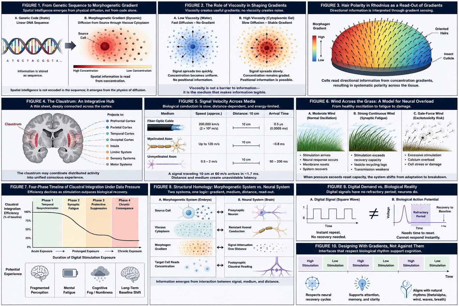
The first conceptual figure appears on page 2. Figure 1 is described as a diagram contrasting a static DNA sequence with a dynamic morphogenetic gradient. The left side shows a linear sequence, while the right side shows a radial diffusion gradient moving from a source cell through viscous cytoplasm. The purpose of this figure is to make the paper’s first major claim visible: spatial intelligence does not simply sit inside the genetic sequence; it emerges when molecular products move through physical space. However, this figure should not be read as experimental data. It is a conceptual diagram designed to clarify the argument.
Figure 2, described on page 3, compares diffusion in water with diffusion in a viscous gel. The low-viscosity example shows rapid, uniform spread, while the viscous example shows slower diffusion and a clearer gradient. This figure supports the manuscript’s claim that resistance can create informational structure. It does not prove a new biological mechanism by itself; rather, it illustrates why Crick’s morphogen model required attention to cytoplasmic viscosity.
The paper then explains morphogens in more concrete biological terms. It states that morphogens are not metaphors; they are physical molecules with measurable properties such as molecular weight, diffusion coefficient, and half-life. Their behavior can be described using diffusion physics, especially the logic of Fick’s laws, which explain how concentration gradients change over time. The key editorial point here is that the manuscript treats biology as material computation. Genetic information does not become form through abstract instruction alone. It becomes form through molecules moving through a resistant medium.
On page 4, the manuscript discusses hair polarity in the cuticle of the insect Rhodnius, drawing on Peter Lawrence’s work as cited through Ridley. The example matters because polarity involves not only what a cell becomes, but which direction it faces. The manuscript explains that hairs on the insect cuticle show systematic orientation, suggesting that cells respond to directional information across a gradient. Figure 3 is described as a schematic of directional hair orientation overlaid with a color gradient. This figure helps readers understand how a visible biological feature, such as hair direction, can become the physical read-out of an invisible chemical gradient. Again, this is used as explanatory support, not as newly produced experimental evidence in DeSilets’ paper.
After establishing this developmental biology framework, the paper makes its major conceptual move: it shifts from embryo to brain. The bridge is the idea that both systems depend on signals moving through physical media. In the embryo, morphogens move through cytoplasmic or extracellular environments. In the brain, electrochemical signals move through neurons, axons, synapses, and distributed neural networks. The paper focuses especially on the claustrum because of its proposed role in integrating signals across the cortex.
The claustrum is described on page 4 as a thin, bilaterally symmetric sheet of gray matter located deep in the brain between the insula and the putamen. The manuscript cites Crick and Koch’s 2005 proposal that the claustrum may function like a conductor, helping coordinate activity across many cortical regions. This is a strong conceptual choice because the claustrum is not presented as a simple sensory area. Its significance comes from connectivity: it is linked with multiple cortical systems, including prefrontal, sensory, visual, auditory, and limbic regions.
Figure 4, described on page 5, shows the claustrum highlighted in a coronal brain section, with radiating lines to cortical regions. This figure supports the paper’s argument that the claustrum can be interpreted as an integrative hub. What it does not prove is that the claustrum is definitively the seat of consciousness or that it alone controls digital overload. The paper relies on an existing theoretical tradition around claustral integration, but its own extension to digital cognition remains speculative and conceptual.
The paper’s next major issue is latency. Digital systems often feel instantaneous to users. Fiber-optic transmission can move signals at enormous speeds relative to biological neural conduction. Human neural signals, by contrast, are electrochemical events. They depend on ion movement, membrane dynamics, axon diameter, myelination, and synaptic transmission. The manuscript gives specific conduction figures: unmyelinated fibers may conduct at roughly 0.5–2 meters per second, while myelinated fibers can reach up to about 120 meters per second. It also gives a concrete timing example: a signal traveling 10 centimeters at 60 meters per second would arrive after approximately 1.7 milliseconds.
That 1.7 millisecond example is important because it shows how small biological delays can still matter. In ordinary language, a millisecond feels negligible. But in neural integration, timing differences can influence whether distributed signals are bound into a coherent perceptual moment. The manuscript argues that consciousness, attention, and perception depend not merely on signal arrival, but on coordinated timing across regions. The claustrum, in this account, does not eliminate the physics of distance; it manages within those limits.
Figure 5, described on page 6, compares signal velocity across three media: fiber-optic cable, myelinated axon, and unmyelinated axon. The figure’s conceptual meaning is clear: digital signal transmission and biological conduction do not share the same temporal physics. Digital systems can approximate near-zero latency at human scales, while biological systems produce distance-dependent arrival times. This figure supports the paper’s biological mismatch argument. It does not, however, directly measure claustral performance under screen exposure; it illustrates the timing difference on which the later theoretical claim depends.
The most vivid part of the manuscript is the “wind across the grass” model. DeSilets uses the metaphor of wind flattening grass and gale-force pressure dispersing dandelion spores to explain neural overload. The paper carefully maps this metaphor onto three biological regimes. Moderate wind corresponds to normal neural oscillation: stimulation arrives, the neuron responds, the membrane resets, and the system becomes ready again. Strong continuous wind corresponds to synaptic fatigue: the system is pressed faster than it can recover. Gale-force wind corresponds to excitotoxicity, where excessive stimulation can contribute to destructive cellular processes.
The strength of this section is that it translates difficult neuroscience concepts into everyday experience without abandoning the biological mechanism. Synaptic fatigue refers to the reduced ability of a synapse to transmit signals during sustained high-frequency activity, often because neurotransmitter vesicle release and recycling cannot keep up. Excitotoxicity refers to a much more severe process in which excessive excitatory signaling, often involving glutamate and calcium influx, can trigger cell damage or death. The paper does not claim that ordinary screen use automatically causes excitotoxic cell death. Rather, it places fatigue, suppression, and structural vulnerability on a conceptual continuum of biological response to excessive pressure.
Figure 6, described on page 6, is a three-panel illustration: moderate wind with recovery, sustained high-velocity wind with flattened grass, and gale-force wind with dandelion spores released. Its scientific value is explanatory and metaphorical. It helps readers visualize three different regimes of biological stress: normal oscillation, synaptic fatigue, and irreversible damage. Its limitation is equally important: it is not an empirical figure generated from neural recordings, imaging data, or clinical measurements.
The paper then applies this model directly to the claustrum under “data pressure.” The term data pressure is used to describe a condition in which the speed, frequency, and simultaneity of incoming stimuli exceed the biological reset capacity of the receiving system. This is where the manuscript becomes most relevant to contemporary digital life. Smartphones, social media feeds, notification systems, multitasking interfaces, streaming platforms, and rapid visual environments can generate continuous multimodal input. The biological receiver, however, still operates through neurons, synapses, refractory periods, and finite biochemical resources.
DeSilets proposes a four-phase model of claustral stress under sustained digital stimulation. Phase 1 is temporal desynchronization. Because different neural signals travel different distances and arrive with different latencies, the claustrum’s integrative role depends on timing alignment. Under excessive input, this alignment may become harder to maintain. The manuscript links this to fragmentation of perception, narrowed attention, and weakening of the unified present moment. This is a theoretical interpretation, not a demonstrated clinical diagnosis.
Phase 2 is synaptic fatigue. In this phase, sustained stimulation causes vesicle recycling and neurotransmitter availability to fall behind demand. The claustrum becomes a bottleneck not because it chooses to filter information intelligently, but because the biological materials needed for transmission are strained. This is one of the paper’s most important claims because it reframes overload as a material limitation rather than a failure of willpower. The problem is not simply that people are distracted; the paper suggests that the receiving tissue may have biological recovery requirements that digital environments ignore.
Phase 3 is protective suppression. The manuscript describes inhibitory mechanisms, including increased GABAergic tone and hyperpolarization, as ways the nervous system may reduce firing under overstimulation. In everyday terms, this may correspond to cognitive fog, numbness, dissociation, or attentional collapse after prolonged exposure to screens. The article must be careful here: the paper does not provide patient data proving that all such symptoms are claustral suppression. It proposes a biologically plausible explanatory framework that would require direct empirical validation.
Phase 4 is chronic structural consequence. The manuscript refers to long-term depression of synaptic weights, reduced dendritic spine density, and receptor expression changes in circuits exposed to chronic overstimulation. This is the strongest and most caution-demanding part of the argument. It links digital overstimulation to possible neural remodeling, but the paper itself does not present longitudinal human neuroimaging, clinical trials, or direct claustrum-specific measurements. Therefore, the claim should be read as a hypothesis-generating theoretical extension, not as settled evidence that digital media chronically restructures the human claustrum.
Figure 7, described across pages 7–8, presents a four-phase timeline of claustral integration efficiency under sustained data pressure. The x-axis moves from acute to chronic digital stimulation exposure; the y-axis represents claustral integration efficiency as a percentage of baseline. The curve begins with a plateau, then declines during synaptic fatigue, stabilizes under protective suppression, and later flattens at a lower chronic baseline. This figure is useful because it organizes the argument into a process model. What it does not prove is the numerical shape of the curve. The paper does not provide empirical measurements of claustral efficiency percentages; the figure is conceptual.
The paper’s central synthesis appears in Section 6, where DeSilets compares morphogenetic systems and neural systems point by point. In both, a signal is produced. In both, the signal travels through a physical medium. In both, the medium has resistance. In both, distance affects signal strength or timing. In both, a receiving element interprets the signal. In both, the system requires reset or recovery. And in both, excessive pressure can collapse the gradient that makes the signal meaningful.
This comparison is the intellectual core of the paper. The embryo and the brain are not being treated as identical, but as structurally analogous systems. In an embryo, if a morphogen floods the field too uniformly, cells lose positional distinctions. In the brain, if digital inputs arrive too continuously and too quickly, the integrative timing that supports coherent experience may degrade. The shared principle is that information does not exist only in the signal. It exists in the relation between signal, medium, distance, timing, and recovery.
Figure 8, described on page 8, places the morphogenetic system and claustral system side by side. The left column shows source cell, viscous cytoplasm, morphogen gradient, and target cell reading concentration. The right column shows presynaptic neuron, resistant axonal conduction, signal attenuation, and postsynaptic claustral reading. This figure supports the manuscript’s claim of “structural homology” between biological signaling systems. The figure is useful for readers because it makes the analogy explicit. Its limitation is that analogy is not identity: morphogen diffusion and neural conduction are different biological processes, and the paper’s bridge between them remains theoretical.
The concept of biological mismatch appears most clearly in Section 6.2. Digital systems, the manuscript argues, have no biological viscosity. They do not need neurotransmitter recovery. They do not experience refractory periods. They can repeat signals indefinitely, distribute stimuli simultaneously, and operate at speeds far beyond the pacing of embodied human experience. Biological cognition cannot do this. The claustrum, and the nervous system more broadly, evolved in a world where stimuli arrived through physical movement, environmental rhythm, distance, and natural intervals of recovery.
Figure 9, described on page 9, compares a digital square-wave signal with a biological action potential curve that includes a refractory trough and recovery to baseline. This is one of the clearest visual expressions of the paper’s claim. The digital system demands another response before the biological curve has reset. The figure does not measure real digital media exposure in users, but it captures a crucial design-theory insight: interface speed may be technically efficient while being biologically misaligned.
The final major contribution of the paper is its design implication. DeSilets does not simply criticize digital technology. She proposes that digital design theory should learn from gradients. A gradient is not less information; it is information structured through distance, attenuation, pacing, and medium sensitivity. If designers treat the human nervous system as a frictionless receiver, they risk producing environments that flatten attention rather than support cognition. If they design with biological viscosity in mind, they may create interfaces that include recovery intervals, temporal gradients, lower-stimulation phases, and rhythms that better respect neural reset.
Figure 10, described on page 10, proposes a temporal interface rhythm with alternating high-stimulation and low-stimulation intervals. The manuscript links this to neural oscillatory rhythms, mentioning theta at 4–8 Hz and alpha at 8–12 Hz, and compares it with natural environmental patterns such as wind gusts and waves. This is a design proposal rather than a tested interface protocol. Its value is conceptual: it encourages designers to think of attention not as an unlimited resource but as a biological process that requires rhythm, recovery, and spacing.
The paper’s strongest contribution is its cross-disciplinary synthesis. It brings together Crick’s work on morphogen gradients, the physics of cytoplasmic viscosity, the claustrum’s proposed integrative role, conduction velocity, synaptic fatigue, excitotoxicity, and digital design theory. It gives readers a powerful language for understanding why more information is not always better information. In biological systems, too much signal delivered too quickly can erase the very gradients that make interpretation possible.
The paper’s weaker point is evidentiary. It is not an empirical neuroscience study. It does not provide original brain imaging data, electrophysiological recordings, clinical assessments, behavioral experiments, or controlled digital-exposure trials. It does not directly demonstrate that the claustrum becomes desynchronized by digital interfaces, nor does it measure synaptic fatigue in humans during screen use. Its claustral data-pressure model is therefore best understood as a theoretical framework that organizes existing biological concepts into a new design hypothesis.
This distinction matters. The paper should not be used to claim that digital technology has been proven to damage the claustrum, cause excitotoxicity, or produce specific clinical disorders. What it does support is a more careful question: if biological cognition depends on timing, recovery, and medium-specific limits, should digital systems be designed as if human users have none of those limits? DeSilets’ answer is no. The human brain is not a fiber-optic cable. It is wetware: electrochemical, temporally constrained, metabolically limited, and dependent on structured recovery.
In historical context, the paper extends a long scientific movement away from viewing biological information as abstract code alone. Crick’s work after the double helix helped show that form arises when molecular signals interact with physical environments. DeSilets applies this lesson to digital culture. Just as an embryo needs a gradient to know position, consciousness may need temporal gradients to maintain coherence. A flattened medium cannot read position. A continuously overstimulated cognitive system may struggle to read time, attention, and experience as a coherent whole.
In present-day terms, the paper speaks to concerns about attention fatigue, multitasking, notification overload, screen exposure, and the design of digital environments. Its argument is especially relevant to human-computer interaction, educational technology, interface design, cognitive ergonomics, architecture, digital media studies, and design ethics. It suggests that the next stage of digital design should not only ask what technology can transmit, but what the human nervous system can responsibly receive.
For the future, the paper points toward testable research directions. Neuroscientists could examine whether sustained multimodal digital stimulation alters claustral-cortical synchronization. Cognitive scientists could measure recovery intervals after high-density screen exposure. Interface designers could compare continuous-feed environments with gradient-based interfaces that include pacing and recovery. Developmental biologists and theorists of morphogenesis may also find in the paper a broader philosophical claim: biological meaning is not produced by code alone, but by code moving through matter.
The final lesson of the paper is both scientific and ethical. Biological systems are not weak because they have friction. They are intelligent because their friction structures information. Viscosity, delay, resistance, and recovery are not defects to be eliminated. They are the conditions that allow living systems to form bodies, coordinate perception, and maintain coherent experience. Digital design that ignores these conditions may become faster while becoming less humane. Design that respects them may become slower in the right way: not less advanced, but more biologically literate.
Source and Method Note
The source analyzed here is The Viscous Code: Morphogenetic Gradients, Neural Latency, and the Biological Mismatch of Digital Cognition by Deborah DeSilets, Doctoral Candidate in the Doctor of Design program at Florida International University, College of Communication, Architecture + The Arts. The manuscript lists Neil Leach as Chair and states that it was submitted to SSRN in May 2026. The PDF also identifies it as an SSRN Working Paper in the Digital Design Theory working paper series.
This source should be treated as a theoretical SSRN working paper and doctoral working manuscript, not as a clearly peer-reviewed journal article. The PDF does not show a journal publication record, acceptance notice, conference proceeding status, or DOI for the paper itself. For that reason, its peer-review status is not clearly peer reviewed. Because it is presented as an SSRN working paper posted for scholarly discussion, its claims should be interpreted as conceptual and hypothesis-generating rather than settled empirical evidence.
The paper’s method is not a laboratory experiment, clinical trial, computational simulation, survey, or statistical meta-analysis. It uses theoretical synthesis and conceptual modeling. It connects Francis Crick’s work on morphogenetic diffusion gradients, the role of cytoplasmic viscosity, Alan Turing’s morphogenesis framework, Peter Lawrence’s polarity observations, Crick and Koch’s claustrum hypothesis, neural conduction velocity, synaptic fatigue, excitotoxicity, and design theory. The manuscript relies on published references to build a cross-disciplinary analogy between morphogenetic signaling and neural integration under digital stimulation.
No original dataset, patient group, neuroimaging cohort, electrophysiological experiment, animal model, or human screen-exposure study is presented in the manuscript. Numerical values discussed in the paper include neural conduction speeds of approximately 0.5–2 meters per second for unmyelinated fibers and up to about 120 meters per second for myelinated fibers, plus the example that a signal traveling 10 centimeters at 60 meters per second would arrive after approximately 1.7 milliseconds. These values are used to support the paper’s latency argument, not to report new experimental findings.
The manuscript contains ten figure descriptions. Figure 1 on page 2 contrasts static genetic sequence with dynamic morphogenetic gradient. Figure 2 on page 3 compares rapid diffusion in water with slower gradient formation in viscous gel. Figure 3 on page 4 illustrates hair polarity as a read-out of a morphogen gradient. Figure 4 on page 5 shows the claustrum as an integrative hub with cortical projections. Figure 5 on page 6 compares fiber-optic, myelinated axon, and unmyelinated axon signal timing across distance. Figure 6 on page 6 explains the wind-grass-dandelion metaphor for normal neural oscillation, synaptic fatigue, and excitotoxicity. Figure 7 across pages 7–8 proposes a four-phase timeline of claustral integration efficiency under data pressure. Figure 8 on page 8 compares morphogenetic and neural signaling systems. Figure 9 on page 9 contrasts digital square-wave demand with biological action-potential recovery. Figure 10 on page 10 proposes a gradient-respecting digital interface rhythm. These figures are conceptual explanatory diagrams, not empirical data plots from new experiments.
Any formulas or physical principles discussed, such as diffusion gradients, Fickian diffusion logic, conduction velocity, latency, refractory periods, and oscillatory frequency bands, are used for explanatory purposes. They should not be read as engineering certification, medical diagnosis, clinical treatment recommendation, safety approval, legal advice, investment advice, policy order, or official regulatory conclusion.
Because the paper is not clearly peer reviewed and appears as SSRN working paper evidence, its conclusions should be handled carefully. Its strongest value is in proposing a design-theory and cognitive-materiality framework. Its claims about claustral desynchronization, synaptic fatigue under digital data pressure, and chronic structural consequences require direct empirical testing before they can be treated as established neuroscience or clinical fact.